
 when an organic fluorophore was in circulation in the mouse. Credit: Zhuoran Ma and Hongjie Dai.")
A new method for analyzing deep-tissue medical images utilizes artificial neural networks to decrease background noise, giving medical practitioners a clearer, sharper image of deep tissues than currently available.
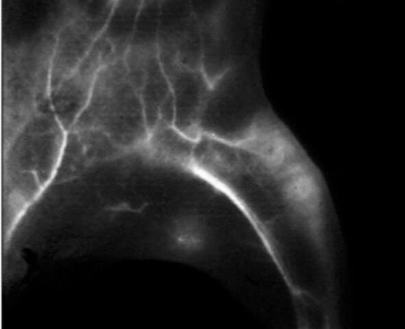
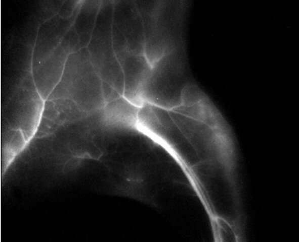
Near-infrared (NIR) fluorescence imaging is a tool used to visualize vascular structures and lymph systems in tissue. For live subjects, the process is called NIR-I, or NIR level one. It works by firing individual protons at an area of the body containing a biocompatible contrast dye, usually indocyanine green (ICG), which then fluoresces and reveals the structure inhabited by the dye. This is useful in tumor growth monitoring, for example, or when diagnosing various cancers.
For nonliving tissue images, a deeper probe called NIR-IIb uses contrast materials containing PbS/CdS quantum dots. NIR-IIb can achieve a much clearer image, but those contrast agents are not approved for human use. The goal for this research, conducted by Zhuoran Ma and a team of researchers from the Stanford laboratory of Dr. Hongjie Dai, was to achieve an image clarity of NIR-IIB using only agents approved for NIR-I procedures, or human-safe, FDA-approved contrast agents.
To reduce light scattering in the biocompatible dyes that cause shallow imaging depth, low contrast and poor clarity in NIR-I images, researchers trained an artificial neural network to remove noise, increase contrast, and ultimately achieve “an image closely resembling the ground truth.” To do so, the artificial neural network was given 2800 in vivo images of mice; the NIR-I images were fed through the network then compared by researchers to NIR-IIb images of the same mice. Through a series of refinements, the artificial neural network was able to improve an NIR-I image to 85% of the quality of an NIR-IIb image.
Adopting this method of image refinement would impact diagnosticians who rely on imaging, particularly for deep tissue diseases such as breast cancer. Current tools allow clear imaging mere nanometers below the surface before scattering begins to blur and distort the structures. Image guided surgeries could also be made simpler to perform and more successful overall if imaging allowed higher contrast and clarity in deep live tissue.
Source: Read Full Article