A team of researchers has studied the molecular profile of small “messenger” vesicles called exosomes, produced by cancer stem cells (CSCs), which play a key role in the process of carcinogenesis and metastasis in the blood of patients with malignant melanoma.
The study has shown that these malignant melanoma vesicles produced by CSCs have a different molecular composition from that of differentiated tumor cells. These molecules were also found to be detectable in exosomes present in the blood, and they presented differences in patients with malignant melanoma compared to healthy individuals. This makes them potentially suitable as biomarkers for the diagnosis and prognosis of this disease.
The results have been published in the journal Molecular Oncology.
Malignant melanoma is one of the most aggressive types of skin cancer and its prevalence has been increasing worldwide in recent years. Among the factors that contribute to the life-threatening nature and severity of this disease are the late appearance of the first symptoms, the lack of effective treatments, its high metastasis capacity, and also the difficulty of detecting this particular cancer. Unfortunately, the diagnosis of malignant melanoma therefore continues to be problematic due to the lack of indicators—known as biomarkers—to accurately signal the early stages of this disease and predict how it might evolve in a given patient, once detected.
These characteristics, which make this type of cancer such a serious disease, may be partly attributable to so-called cancer stem cells (CSCs), a sub-population of cells that exist in tumors and that present the typical characteristics of stem cells. They are responsible for tumor initiation, maintenance, and progression, as well as metastasis and recurrence—even years after a tumor has been eradicated.
Now, a team of scientists led by Professor Juan Antonio Marchal Corrales of the Department of Human Anatomy and Embryology at the University of Granada (UGR) and Director of the “Doctores Galera y Requena” Chair in Research on Cancer Stem Cells, pertaining to the Biohealth Research Institute in Granada (ibs.GRANADA) and the MNat Scientific Unit of Excellence (ModelingNature), has studied these CSCs—specifically, the microvesicles that act as “messengers” for these cells. Known as exosomes, these cells produce and send other cells and tissues to communicate via the transfer of certain biomolecules, thereby promoting the emergence of metastases.
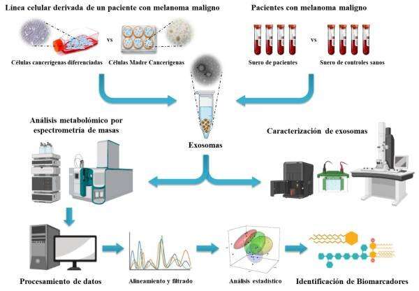
These exosomes have been shown to be involved in many tumor processes. As cells release them and circulate via the bloodstream, they offer a very interesting source of biomarkers as they can be easily isolated from a blood sample. This study focused on the molecular characterization of exosomes produced by CSCs and isolated in the blood from patients with malignant melanoma. Metabolomic techniques were used to analyze the molecular profile of biological systems in order to identify possible biomarkers for the diagnosis of this disease.
This study is the result of extensive multidisciplinary work in which translational researchers, bioinformaticians, and clinical researchers have joined forces to take another step in the field of Personalized Medicine or Precision Medicine in Oncology. The team comprises members from the UGR; Fundación MEDINA (led by Francisca Vicente and José Pérez del Palacio, Area Head and Principal Investigator of the Screening Department, respectively); and the “Virgen de las Nieves” and “San Cecilio” Teaching Hospitals in Granada (all members of ibs.GRANADA ); the University of Vigo; and the Spanish National Cancer Research Centre (CNIO).
Among its findings, the study showed that the molecular composition of exosomes produced by CSCs is different from those released by differentiated tumor cells. To investigate this, using a primary patient-derived malignant melanoma cell line enriched in CSCs, both types of cells were cultivated in large quantities and the exosomes that they produced and released into the culture were isolated. Once the properties and characteristics of both the cells and the exosomes they produced had been tested, a metabolomic analysis was carried out. This enabled the molecules (metabolites) present in the biological sample to be studied. After the molecules had been detected and extracted using a mass spectrometer, which quantifies them with great precision, a series of statistical analyses were carried out to determine which molecules were found in the highest concentration in the exosomes of each cell type. Thus, the researchers tentatively identified some lipidic metabolites differentially present in exosomes of CSCs and differentiated tumor cells.
Metabolomic profile
Subsequently, and following the same scientific approach, a similar study was carried out comparing the metabolomic profile of exosomes isolated from the blood of patients with malignant melanoma in different stages and healthy individuals who acted as controls. The study concluded that certain metabolites, including some of those previously identified in CSCs, were also present in exosomes isolated from blood in different concentrations among melanoma patients and healthy individuals. By means of the corresponding statistical models, these molecules and their different concentrations in blood made it possible to distinguish individuals with malignant melanoma from those without the disease. This makes them suitable candidates for acting as potential biomarkers for its diagnosis.
However, the authors emphasize that this study is only a first step. The identification of some of these molecules, the complete characterization of those already tentatively identified, and the replication of the study with a greater number of samples to validate and verify their clinical application as biomarkers all remain pending.
Source: Read Full Article
