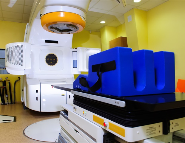
Patients with cancer that has spread to the bone are sometimes treated with alpha particle radiation therapy that is delivered into or very close to the tumor. However, it is unknown whether the radioactive particles are distributed through the surrounding region or to the body's vital organs, where they could have toxic effects.
Abhinav Jha, an assistant professor of biomedical engineering at the McKelvey School of Engineering and of radiology at the School of Medicine's Mallinckrodt Institute of Radiology (MIR), both at Washington University in St. Louis, and colleagues plan to measure this distribution using a novel imaging method with a four-year $2.2 million grant from the National Institutes of Health (NIH).
Using a novel low-count quantitative single photon emission computed tomography (LC-QSPECT), Jha plans to build a computational framework from which to measure the concentration of the radiopharmaceutical material. Scans with this technology will allow the team to measure the concentration of the radiopharmaceutical activity in the tumor and the various radio-sensitive organs of the body.
Understanding the distribution of the drug inside the body helps plan the treatment. Also, one key concern is underdosing, which would not kill the tumor."
Abhinav Jha, Assistant Professor of Biomedical Engineering, McKelvey School of Engineering and of rRadiology at the School of Medicine's Mallinckrodt Institute of Radiology
SPECT imaging could provide a mechanism to see where the drug has gone in the body. However, the challenge is that the number of counts detected with these treatments is very small. Conventional approaches that reconstruct the distribution of the isotopes and estimate the uptake from reconstructed images are not accurate at low count levels, so there is a need for new approaches to quantify the drug in a patient.
Drug Discovery eBook

The approach that Jha and colleagues propose stems from previous research published earlier this year in IEEE Transactions on Radiation and Plasma Sciences. In that research, they found that a low-count quantitative single-photon emission computed tomography (LC-QSPECT) method provided reliable measurements of the radionuclide uptake.
To validate this method, Jha and his team also plan a human trial in patients with metastatic prostate cancer that no longer responds to hormone therapy, or castrate resistant, to validate this method. The team has already done a computational clinical trial in a simulated patient population. The results showed that the method was yielding highly accurate and precise values of radionuclide organ uptake.
"Our eventual goal is clinical translation of this method so that it can benefit patients being treated with these therapies." Jha said. "There is much excitement surrounding this use of SPECT imaging for therapy. We are delighted to have the opportunity to contribute to this space."
Jha will be joined in these efforts by faculty from the School of Medicine, including Brian Baumann, MD, an assistant professor of radiation oncology, and the following faculty from the MIR: Daniel L.J. Thorek, an associate professor of radiology; Richard Laforest, a professor of radiology; Farrokh Dehdashti, MD, a professor of radiology and senior vice chair and division director of nuclear medicine; Tyler J. Fraum, MD, an assistant professor of radiology; and Richard L. Wahl, MD, the Elizabeth E. Mallinckrodt Professor of Radiology and MIR director.
Washington University in St. Louis
Posted in: Device / Technology News | Medical Science News
Tags: Bone, Cancer, Catalyst, Clinical Trial, Computed Tomography, Education, Hormone, Imaging, Medicine, Nuclear Medicine, Oncology, Prostate, Prostate Cancer, Radiation Therapy, Radiology, Radionuclide, Research, students, Tomography, Translation, Tumor
Source: Read Full Article
