A review paper by scientists at the Chonnam National University summarized the recent research on bioprinting methods for fabricating bioengineered blood vessel models. The new review paper, published in the journal Cyborg and Bionic Systems, provided an overview on the 3D bioprinting methods for fabricating bioengineered blood vessel models and described possible advancements from tubular to vascular models.
“3D bioprinting technology provides a more precise and effective means for investigating biological processes and developing new treatments than traditional 2D cell cultures. Therefore, it is a crucial tool in the field of regenerative medicine and biological research,” explained study author Hee-Gyeong Yi, a professor at the Chonnam National University.
Two-dimensional (2D) blood vessel models have difficulties in mimicking the three-dimensional (3D) microenvironment in human, simulating kinetics related to cell activities, and replicating human pathophysiology.
“In vitro bioengineered models created through biofabrication based on tissue engineering and regenerative medicine are breakthrough models that can overcome limitations of 2D and animal models,” said study authors. Thus, they reviewed the 3D bioprinting methods for fabricating bioengineered blood vessel models and described possible advancements from tubular to vascular models.
Bioengineered models (BMs) are innovative models that can overcome limitations of 2D and animal models to allow simulation of the natural microenvironment in the human body in a patient- and target-specific manner. “BMs can be used to verify the safety and efficacy of drugs and medical devices by recreating structures and functions that maximally resemble those of tissues and organs in vitro,” said Yi.
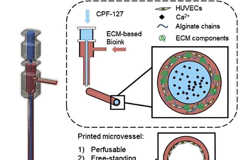
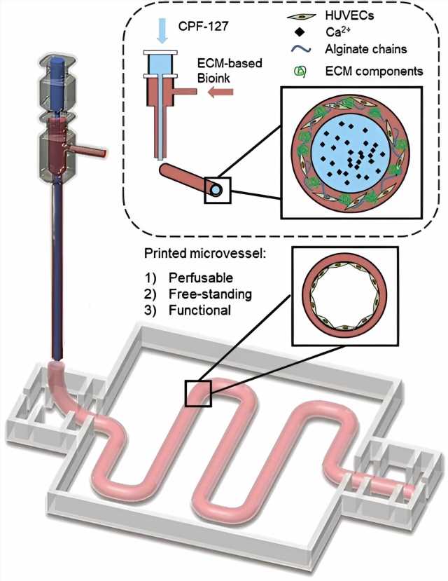
The study authors discussed recent 3D bioprinting methods for fabricating bioengineered blood vessel models. Coaxial nozzle bioprinting, for example, is a 3D bioprinting technique used to fabricate structures of concentric shapes via simultaneous printing of bioink of two different materials.
Bioprinting can be used to fabricate blood vessels with complex, micro-scale structures in vitro for the construction of in vitro models featuring multiple connected tissues. Moreover, fabrication of vascularized tissues that closely resemble anatomical tissues will allow a detailed in vitro examination of diseases related to blood vessels and further large-scale fabrication of diverse, large-caliber tissues.
Recent rapid advancement of techniques to fabricate in vitro models is expected to overcome current limitations, paving the way for more accurate drug evaluation and efficacy analyses of blood vessels and blood flow dynamics in the body.
“Advancements in scaffold fabrication techniques, such as electrospinning or 3D scaffolding approaches, and fine-tuning printing parameters based on the specific requirements of tubular structures and complex tissue models can contribute to the creation of more intricate and tailored structures,” said Yi.
The review paper calls for researchers, medical professionals, engineers, and other experts to collaboratively marshal the research on 3D bioprinting methods for fabricating bioengineered blood vessel models into practical applications that study human physiology and diseases in a more relevant and accurate manner.
More information:
Seon-Jin Kim et al, Bioprinting Methods for Fabricating In Vitro Tubular Blood Vessel Models, Cyborg and Bionic Systems (2023). DOI: 10.34133/cbsystems.0043
Journal information:
Cyborg and Bionic Systems
Source: Read Full Article
