Biopsies haven’t changed much in the more than 100 years they’ve been in medical use: Tissue (a cluster of cells) is cut from the body, embedded into a block, thinly sliced, mounted on a slide, and stained with dye. A pathologist then analyzes the sample with a microscope. Results come back in 2 to 10 days while the patient and their family nervously wait.
Engineers at Columbia University are working to give biopsies a much-needed upgrade. There, Elizabeth Hillman, PhD, and her team have developed a high-speed 3D microscope that can rapidly take photos of live cells without having to extract them from the body.
The result: A noninvasive approach where results happen a whole lot faster.
Medical Imaging Meets ‘The Matrix’
We already use microscopes in some surgeries, but most provide only a small 2D picture, limiting the view of important details.
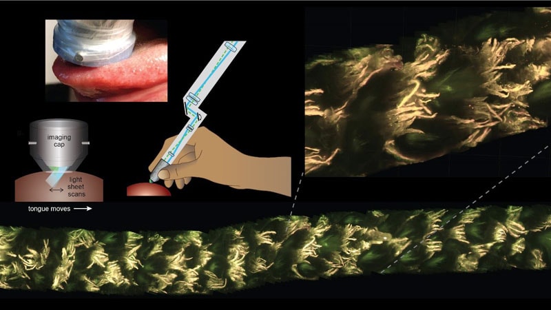
“Tissue looks different at different depths,” Hillman says. “If you have a 3D image of the tissue, you can look at it at different levels — something that can’t be done with 2D images.”
Until now, examining those depths required a scalpel. But the technology Hillman and her team have developed, called MediSCAPE, involves simply dragging a small probe across the tissue. The probe very quickly takes many pictures of the living cells, making a large-scale 3D view of the tissue’s tiny features.
The difference between a 2D scan and the 3D version is a bit like comparing a flat Polaroid picture to the “Bullet Time” scene from The Matrix. High-speed imagery from many different angles offers a level of detail and precision that a 2D picture can’t capture.
This clearer picture lets surgeons better tell healthy tissues from unhealthy ones, letting them decide how best to cut out a tumor so no diseased tissue remains. Best of all, if the image looks normal, the tissue gets to stay where it is, inside the patient.
The technology could be useful not just for spotting tumors, but also “in guiding surgeons by quickly identifying different types of tissue like nerve, fat, muscle, cartilage, scar tissue,” Hillman says.
Are Better, Safer Biopsies Ahead?
Hillman cautions that high-speed 3D imaging is not intended to replace all biopsies. But MediSCAPE could be especially useful in examining sensitive areas like the brain, where removal of such precious tissue might lead to loss of function as well as swelling, seizures, or strokes.
Early uses for the technology will most likely be in open surgeries where the patient’s tissues are exposed — such as in the brain, abdomen, cervix, ear/nose/throat — and to guide complex robotic surgeries, she says.
Still, while the study proves the technique is possible, it could be another 5 years or more before the technology is available mainstream, Hillman says. Many more clinical trials lay ahead.
Sources:
Elizabeth Hillman, PhD, professor of biomedical engineering and radiology (physics), Columbia University.
Nature Biomedical Engineering: “High-speed light-sheet microscopy for the in-situ acquisition of volumetric histological images of living tissue.”
Source: Read Full Article