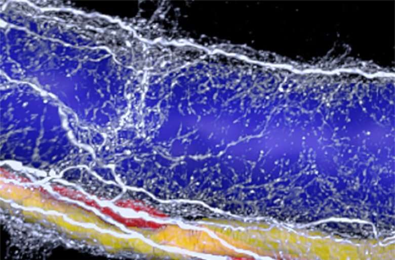
With the application of a novel three-dimensional imaging technology, researchers at Karolinska Institutet have discovered that one portion of the autonomic nervous system in the liver undergoes severe degeneration in non-alcoholic fatty liver disease. The study, which is conducted in mice and human liver tissue, shows that the degeneration of nerves is correlated with the severity of liver pathology. The results are being published in the journal Science Advances.
Non-alcoholic fatty liver disease is the most common hepatic disorder, with prevalence around 25 percent globally. Approximately one third of all fatty liver cases will develop to steatohepatitis, which is a severe disease seriously affecting the entire metabolism.
In the current paper, researchers explore the nervous system in fatty liver using volume immuno-imaging and light sheet microscopy ― a novel imaging technique, which altogether offer large-scale 3D visualization with cellular resolution. According to the study, this technology can reveal even early, minor or hidden structural impairments of the liver.
“Now we know that nerves in the liver have multiple subtle regulatory roles” says Csaba Adori, researcher at the Department of Neuroscience, who led the study. “Their role, however, may be more essential during the fight-or-flight response or when subjected to metabolic challenges. Degeneration of liver sympathetic nerves and abnormal operation of the remaining nerve fibers in the fatty liver could compromise all these functions, which may contribute to further aggravation of the disease, as part of a vicious cycle.”
Occurs already in early stages
Source: Read Full Article
