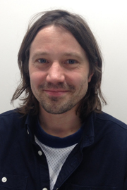
 Thought LeadersSerge Mostowy and Sydney MilesLondon School of Hygiene and Tropical Medicine
In this interview, NewsMedical speaks with Professor Serge Mostowy and Sydney Miles about their research on Shigella Flexneri and enteropathogens. They discuss how new automated approaches are enabling new insights, never before achievable. In particular, they focus on an automated, AI-powered analysis software, Athena, that is available for download in a novel pay-per-use basis.
Could you please introduce yourself, what it is you do and briefly describe your research?
Serge Mostowy: My name is Serge Mostowy and I am a Professor of Cellular Microbiology at the London School of Hygiene and Tropical Medicine, located in central London.
In the laboratory, we focus on new ways to control bacterial infection. Our bacterial pathogen of focus is Shigella flexneri. It is a very important human pathogen, responsible for hundreds of millions of illness episodes per annum, but it is also a paradigm of discovery in the innate immune response, historically having enabled many discoveries in this field.
We feel that if we continue to use Shigella, we can continue to discover amazing things. To make a translational impact, we focus on going from the single, infectious bacterial cell to the consequence over the whole animal. Therefore, we utilize zebrafish larvae to understand the whole animal response to Shigella infection.
Sydney Miles: My name is Sydney and I am a Ph.D. student in the lab of Serge Mostowy at the London School of Hygiene and Tropical Medicine. My project focuses on investigating the evolution of enteropathogens. This essentially involves collecting a bunch of strains of different backgrounds and we infect zebrafish with them to try and understand what makes one strain better than the other.
We look for differences in host response, such as how the number of neutrophils and macrophages in a zebrafish changes over time. This will help us understand why some strains are able to infect humans better than others.
What are the major challenges you face in getting the information of the samples that you need?
Serge Mostowy: Cell biology of infection or cellular microbiology is synonymous with advances in microscopy techniques. New microscopes are evolving faster than iPhones. It is really an amazing time to visualize the infection process at a resolution that was just previously not possible. To visualize infection from the level of the single cell to the whole animal, we use the zebrafish larva.
In terms of challenges, following the cells or the infection process at a resolution not possible to achieve in other labs, requires the highest end-resolution microscopes. It also requires better tools to analyze and understand the infection process.
Not only are you annotating and characterizing the type, site and dissemination of the infection, but quantifying both bacterial replication, whole animal survival and host response is also important. As microscopes continue to evolve, better tools are required to understand and quantify the infection process.
What advantages does a tool like the Athena software have over other more conventional methods?
Serge Mostowy: It is an exciting time because tools such as the Athena software are enabling us to quantify parameters at a pace and resolution that was not previously achievable and that had to be manually quantified previously.
Now, with tools like Athena, this process is completely automatic, enabling more high-throughput analysis techniques and eliminating user bias. Such developments have been transformative for my lab, especially the speed at which we can do things now.
Sydney Miles: Running studies for 60 different fish at a time was really challenging and it would often take hours to sit there and count manually. However, the introduction of Athena has significantly accelerated the progress that we are able to make.
When you started working on the Athena software, how long did it take you to be independent and productive with it?
Sydney Miles: The first time I opened the Athena software, I had IDEA Bio-Medical on a video call with me, and they ran through it with me, demonstrating how to use the program. From there, it took me about a week to become fully independent.
In a typical experiment, I will use a 96-well plate, so I could put up to 96 fish in there. In my experiments, I analyze 60 fish [in a 96-well plate] in three separate experiments per week, which becomes hundreds of fish per month.
Obviously, counting them would take a really long time, limiting the time I had to perform the experiments. Athena has accelerated the pace that I can work.
 Image credit: IDEA Bio-Medical
At what point did you move away from manual imaging and image analysis to search for automated solutions?
Serge Mostowy: I was fortunate enough to have earned an ERC consolidator grant, allowing us to purchase a high-content, high-throughput microscope, which was a brand-new research direction for my lab. We were then able to move away from the infection of, say, half a dozen to a dozen larvae at a time, to work with 96-well plates.
However, with these different microscopes, you need the innovation of analytical tools and that is where Athena helps us because although we were generating data at a much faster pace, it was no longer feasible for students to quantify manually. It simply takes up too much time.
Now, we prepare and collect data over two or three days because all the analysis is live, and then we plug these images into the Athena software.
With just a few clicks of a button, we can now analyze our areas of interest in the zebrafish larvae and reliably quantify things at a scale that was previously unimaginable. We were able to maximize the time away from the bench using these new software approaches.
What has this technology brought to your lab?
Serge Mostowy: Athena has transformed our ambition. We have research questions that we have received funding to try to answer, but now we can do things on a much larger scale. So instead of some of our graphs where we quantify a dozen or two dozen zebrafish, we can now go to the order of hundreds or even thousands.
The field of zebrafish infection characteristically relies on things like zebrafish survival or bacterial burdens using older techniques. Now with Athena, live larva analysis is possible over time at a much higher resolution, amplifying our potential for discovery.
Things can happen at a much faster pace, and the quantifications are much more reliable. The errors are reduced and I am no longer concerned about human-to-human variation in the analyzed data because this software eliminates this bias.
Overall, our ambition for discovery has been enhanced because we can achieve more, and the quantifications are more reliable. It has added a new dimension to our research.
It is easy to implement the software to count different cell types or the different sites of infection in the fish. So, just because the fish lends itself to microscopy, Athena has really recognized its ability to contribute to novel discoveries.
Sydney Miles: The support is excellent – I have a direct line of contact with IDEA Bio-Medical regarding Athena and that is super helpful.
The main thing I use Athena for is counting individual cells within the whole fish. The feature of setting the parameters once and then going back to it and using that same ones for each and every experiment has radically changed things for me for the better.
Where do you see your work going next, and what is the impact?
Serge Mostowy: I think about this question a lot. We would like to push the software to its limit and help drive future innovation that could really benefit the zebrafish community as a whole.
As we publish our research while we help to develop the software, I think it will make for a more robust science across the entire community.
I also really want to maximize the ambition of our science. As I previously mentioned, we are now going to 96 well plates. We now have the capacity to screen 12,000 compound libraries to maximize the use of the zebrafish as an infection model to bring us closer to understanding human disease. As the microscopes improve, as the software to analyze the microscope images gets better, so does our ability to translate our findings from the fish to one day toward a better understanding of human disease.

About Serge Mostowy, PhD
Prof. Mostowy is a Lister Institute of Preventive Medicine, Wellcome Beit and Wellcome Trust Senior Research Fellow. He studied Physics, Evolution, and Microbiology & Immunology at McGill University in Canada. After postdoctoral work on the Cell Biology of Infection at Institut Pasteur in France, He moved to Imperial College London in 2012 to start a Wellcome Trust Research Career Development Fellowship. In 2018, Prof. Mostowy was appointed Professor of Cellular Microbiology at the London School of Hygiene & Tropical Medicine, where he investigates novel roles for the cytoskeleton in innate immunity. The Mostowy group also developed the Zebrafish as an important model to study the cell biology of infection and therapeutic potential of targeting the cytoskeleton in vivo. Their findings should be of considerable interest to both cell biologists and infection biologists, and make a cutting-edge platform for in vivo studies both at the single-cell and whole-animal level.
Email: [email protected]
Website: www.themostowylab.org Twitter: @MostowyLab
 About Sydney Leigh-Miles
Sydney is currently in the second year of her PhD at the London School of Hygiene and Tropical Medicine, funded by a BBSRC London Interdisciplinary Doctoral Training Programme Studentship. Sydney’s research aims to study the evolution of a globally important human pathogen, Shigella sonnei. I use a combination of microbial genomics and zebrafish infection techniques, ultimately aiming to understand what drives the epidemiological success of some bacterial strains over others.
Twitter: @sydneylmiles
About IDEA Bio-Medical Ltd.
IDEA Bio-Medical is a company specializing in automated microscopy and image analysis for the life science researchers. It was founded in 2007 through a partnership between YEDA (the Weizmann Institute’s commercialization arm) and IDEA Machine Development Design and Production Ltd (an innovation hub). IDEA’s products, the Hermes imaging system and Athena image analysis SW, have contributed to over 100 scientific publications in peer-reviewed magazines, globally supporting high impact science.
IDEA Bio-Medical currently focuses on empowering zebrafish researchers, specifically, to provide them with a reliable, robust solution for automated and unbiased Zebrafish image analysis by applying company’s knowledge and expertise.
To this end, IDEA developed a novel deep learning-based image analysis software for in vivo zebrafish experiments. The software automatically detects zebrafish contour and their internal organs in brightfield with no required user input. The anatomy identified is coupled to fluorescence channels to permit anatomy-specific study of fluorescence changes. It is an affordable, user-friendly system designed specifically for reliable, automated zebrafish image-based analysis.
The software is available as a stand-alone product and accepts microscopy images in multiple image formats, including proprietary ones. It is suited for researchers who only image and analyze a handful of fish per week, as well as researchers imaging hundreds and thousands of fish in multi-well plates for large scale screens. IDEA Bio-Medical is offering a novel pay-per-use model to access the software to enable flexible access. So, all researchers using manual microscopes or automated systems from other vendors, can readily use IDEA’s Zfish software to extract quantitative, meaningful information from their Zebrafish images when they need it.
Reach out to IDEA Bio-Medical on our website's contact form, and more information can be found on the zebrafish analysis software product page.
Sponsored Content Policy: News-Medical.net publishes articles and related content that may be derived from sources where we have existing commercial relationships, provided such content adds value to the core editorial ethos of News-Medical.Net which is to educate and inform site visitors interested in medical research, science, medical devices and treatments.
