Non-alcoholic fatty liver disease (NAFLD) is among the most prevalent chronic liver disorders worldwide and can sometimes lead to severe conditions like cirrhosis and hepatocellular carcinoma. As such, early assessment of the severity of NAFLD is essential for timely intervention. Non-alcoholic steatohepatitis (NASH) and liver fibrosis are two important factors that determine NAFLD progression and probability of cirrhosis development, respectively. So far, liver biopsy has been the most widely recognized method for diagnosing and evaluating NASH and fibrosis. However, it is an invasive procedure that is susceptible to observer bias and suboptimal standardization.
Consequently, recent studies have focused on exploring non-invasive tests for NAFLD, NASH, and fibrosis, for clinical applications. Now, researchers from China have collated recent developments in NAFLD assessment and analyzed the benefits and limitations of the new methods in a review published in the Chinese Medical Journal.
“Accumulating evidence points at different non-invasive tests for diagnosing NAFLD, assessing its severity, and predicting its prognosis. We reviewed the recent literature and summarized the key features of each test,” explains Prof. Vincent Wai-Sun Wong, the corresponding author of this study.
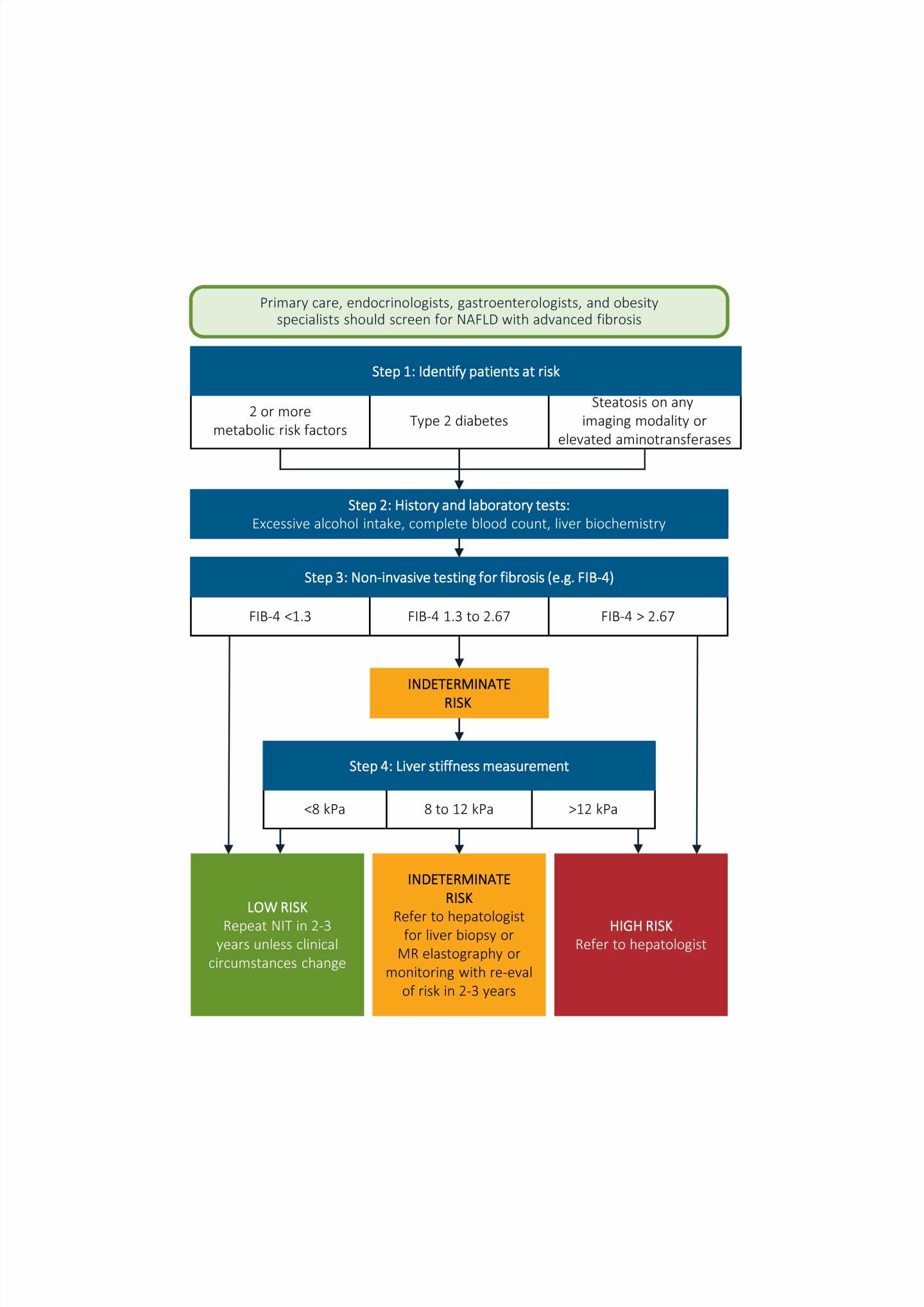
The team clarifies there are two major types of non-invasive tests—blood-based biomarker tests, and imaging methods. Blood-based tests, with multi-biomarker panels, can measure and evaluate biological processes in the liver with decent accuracy. They can be useful for initial diagnosis of liver disorders, since they are more accessible and economic as compared to imaging methods. For example, Fibrosis-4 index and enhanced liver fibrosis panel are promising biomarker tests for detecting advanced fibrosis and predicting its progression. However, some of these tests are influenced by age and gender and have limited efficacy in staging liver disorders.
Imaging methods have proven more accurate in detecting and assessing the severity of liver disorders. For instance, magnetic resonance imaging proton density fat fraction detects NAFLD and NASH with high accuracy, and also stratifies NASH severity. Similarly, machine learning-based ultrasound imaging is gaining popularity for effectively detecting and quantifying NAFLD. Imaging techniques like transient elastography, acoustic radiation force impulse, and magnetic resonance elastography can accurately measure liver stiffness, which is an indicator of fibrosis. However, these methods are often expensive, have limited availability, lack widespread validation, and may require experienced operators.
“Ultimately the selection of appropriate tests for assessing liver disorders is contextual. Availability, cost, and local expertise are key factors to consider while establishing a clinical care pathway for NAFLD,” observes Dr. Wong.
Source: Read Full Article
