
Intravital microscopy is a powerful imaging tool for visualizing various biological processes, simultaneously. This allows for in vivo study of several critical cell types that can be difficult to fully understand when present in cell cultures, such as neurons or tumor cells.
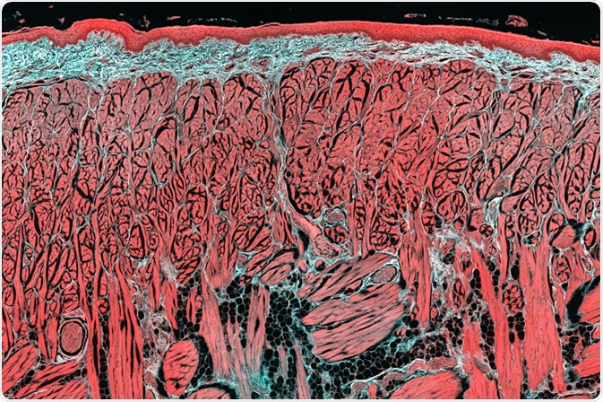 Image Credit: Micha Weber / Shutterstock.com
How does intravital microscopy work?
Intravital microscopy is a term that encompasses several optical microscopy techniques that are applied to living animals for kinetic and functional studies. Its development has been greatly aided by the development of ‘non-linear’ excitation-detecting microscopes.
Non-linear excitation involves the generation of contrast by higher-order interactions of light and biological material. This means that there is usually absorption or scattering and recombination of two photons or more by the specimen when hit by incident light.
One of the forms of intravital microscopy, called multi-photon microscopy, allows for imaging of tissues deeper than that allowed by confocal microscopy. The light source is typically near-infrared or infrared light, which is less significantly scattered or absorbed by biological tissues compared to visible light.
The near-infrared or infrared light is used on the specimen in short pulses of around 100 fs at high rates of repetition (80-100 MHz). In order to achieve the multi-photon excitation, the light needs to be focused directly on the excitation spot. This is done using high numerical aperture lenses.
Another method of intravital microscopy is second and third harmonic generation microscopy, where there is no energy absorption and instead the photons are scattered and recombine into a single photon. This type of method can be applied to tissue that can generate harmonic signals, which include collagen, microtubules, and muscle myosin.
Fluorescence lifetime imaging microscopy is slightly different from the other methods because it relies on the length of a molecules’ excited state rather than photon scatter and recombination. The lifetime measure is different depending on the molecule in focus and is not affected by the molecule’s concentration.
However, it is sensitive to changes in the environment, which means fluorescence lifetime imaging is most commonly used to understand environmental factors such as pH, oxygen levels, and metabolic state of a biomolecule.
Benefits of intravital microscopy
A lot of the knowledge of cellular biology comes from studies on cell cultures, which are very informative for experimental manipulation but are not often good representations of the biological complexity of many cells. Intravital microscopy allows for imaging of live animals, such as the common model organisms C. elegans and D. melongaster.
This allows for understanding complex biological systems in easily manipulated animals, allowing us to understand more about neurobiology, immunology, and cancer.
In addition to its in vivo benefits, intravital microscopy has better penetration than most other microscopy methods. For example, confocal microscopy of biological specimens allows for imaging up to 50-60 µm beneath the surface, whereas multi-photon microscopy (one of the principal forms of intravital microscopy) can reach depths of up to 1 mm.
Thanks to the methods used to focus the light, which means the absorption and emission occur in small volumes, both phototoxicity and photobleaching are significantly reduced. Second and third harmonic generation microscopy are especially good at this, as there is no energy loss in the photon recombination process. The focusing methods also reduce emissions from surrounding tissue that is not in focus.
Applications
Different types of intravital microscopy may be better suited to one application than others. For example, second and third harmonic generation microscopy are best suited for muscle tissue as these have cell types that can produce harmonic signals. However, some evidence shows that lipid bodies can also be imaged using third harmonic generation microscopy.
In cases where there is not necessarily only one method that works, several intravital microscopy techniques can be combined. Multi-photon microscopy is often used in combination with second and third harmonic generation microscopy.
Similarly, fluorescence lifetime imaging can be used with multi-photon excitation and harmonic generation to enable deep tissue imaging. Other types of intravital microscopy that can be used in various combinations include coherent anti-stokes Raman microscopy and optical frequency domain imaging.
Sources
- Masedunskas, A. et al. (2012). Intravital microscopy: a practical guide on imaging intracellular structures in live animals. Bioarchitecture. https://doi.org/10.4161/bioa.21758
- Weigert, R. et al. (2010). Intravital microscopy: a novel tool to study cell biology in living animals. Histochemistry and Cell Biology. https://doi.org/10.1007/s00418-010-0692-z
Further Reading
- All Microscopy Content
- Advances in Fluorescence Microscopy
- Applications in Light Microscopy
- Electron Microscopy: An Overview
- Brief History of Microscopy
Last Updated: Jun 4, 2020

Written by
Sara Ryding
Sara is a passionate life sciences writer who specializes in zoology and ornithology. She is currently completing a Ph.D. at Deakin University in Australia which focuses on how the beaks of birds change with global warming.
Source: Read Full Article