
Fluorescent microscopy has revolutionized the study of bacteria and helped scientists to understand various aspects of their growth, development, and pathogenesis.
Jeerasak Meeraka | Shutterstock
Bacterial cells are around 1 micron in size, which makes them invisible to the naked eye. Fluorescence microscopy allows different parts and aspects of bacteria to be visualized – including nuclei, cell membrane, organelles, and even specific proteins.
Using fluorescence microscopy to study cell numbers
Fluorescent microscopy is often used to visualize nuclei stained using DAPI (4′,6-diamidino-2-phenylindole). This is a DNA stain that appears blue under the microcope. The luminosity of the fluorescent signal from DAPI can increase almost 20-fold upon binding to the AT regions of double stranded DNA.
The excitation wavelength of the stain is in the violet region of the spectrum (i.e. 405 nm). Although it is conventionally used to stain fixed cells, it can also be used to stain the nuclei of live cells when the concentration is increased. Once the nuclei are stained, they can be counted post-imaging using image processing software to count the number of cells present in a sample.
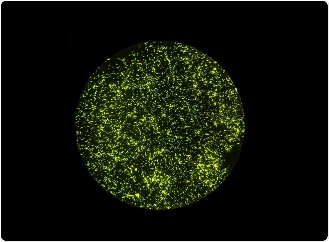
Using fluorescence microscopy to study bacterial cell viability
Fluorescence microscopy can also be used to study the viability of bacterial cells. Fluorescent dyes – such as propidium iodide and SYTO9 – can be used for this purpose. SYTO9 can permeate the membranes and thus can identify all the bacterial population, while propidium iodide is membrane-impermeable.
Propidium iodide is used as a marker of degraded or comprised membranes, thus SYTO9 can be used to differentiate nonviable (dead) bacterial cells from live cells.
Using fluorescence microscopy to study pathogenesis in bacteria
Advances in fluorescent techniques have enabled the visualization of pathogenic and non-pathogenic interaction between host and bacteria.
Fluorescent protein chimeras are fusion proteins that link fluorescent proteins to the bacterial protein of interest. For example, the BCG vaccine acts against Mycobacterium tuberculosis by arresting the vacuolar maturation and preventing phagosome-lysosome maturation. This process can be visualized using fluorescent protein chimeras of Rab proteins that are involved in membrane tracking, including vesicle formation and movement.
Fluorescent probes can also be used to measure pH. Bacterial pathogenesis often results in changes in pH and intracellular free calcium. These parameters can be measured using fluorescent reporters of pH and calcium ions.
Fluorescent probes should be such that low concentrations of the probe can detect minor changes and concentration of intracellular pH and calcium, as the use of high levels of the probe may cause toxicity in targeted cells.
Pseudomonas aeruginosa releasing eDNA, captured by fluorescence microscopy and phase-contrast.
Using fluorescence microscopy to study signaling in bacteria
Different methods are employed to study signaling and protein dynamics in bacterial cells. These methods include single particle tracking (SPT), fluorescence recovery after photobleaching (FRAP), and Förster resonance energy transfer (FRET).
Single particle tracking (SPT) involves imaging and tracking the movement of single fluorescent molecules. For this method to achieve a high signal to noise ratio, the concentration of the fluorescent molecules should be low. Also, the acquisition time should be high as the freely diffusing single molecules travel at a very fast rate.
Fluorescence recovery after photobleaching (FRAP) can be used to measure the movement of molecules inside cells and membrane. In this method, a certain region of the cell is completely bleached. Then the recovery dynamics of the fluorescent protein is captured and measured. This can be used to give an idea of whether a protein molecule is in a free or bound state, and also about the recycling of proteins.
FRET or Förster resonance energy transfer gives a measure of how closely two proteins are located inside cells. The proximity of two proteins is used to infer if there is any signaling between them. Thus, both of these methods help to identify and discover signaling pathways inside bacterial cells.
Sources:
- https://www.ncbi.nlm.nih.gov/pmc/articles/PMC3814296/
- www.microbiology.columbia.edu/faculty/pdf/meyeranddworkin2007.pdf
- pdfs.semanticscholar.org/95a4/6ec5c3b212d8a662570da06f914103e8e51c.pdf
Further Reading
- All Fluorescence Content
- What is Fluorescence Spectroscopy?
- How do Epifluorescence Microscopes Work?
- GFP-tagging in Fluorescence Microscopy
- Fluorescence Quenching
Last Updated: Nov 21, 2018

Written by
Dr. Surat P
Dr. Surat graduated with a Ph.D. in Cell Biology and Mechanobiology from the Tata Institute of Fundamental Research (Mumbai, India) in 2016. Prior to her Ph.D., Surat studied for a Bachelor of Science (B.Sc.) degree in Zoology, during which she was the recipient of anIndian Academy of SciencesSummer Fellowship to study the proteins involved in AIDs. She produces feature articles on a wide range of topics, such as medical ethics, data manipulation, pseudoscience and superstition, education, and human evolution. She is passionate about science communication and writes articles covering all areas of the life sciences.
Source: Read Full Article