Infection with the severe acute respiratory syndrome coronavirus 2 (SARS-CoV-2) that led to the coronavirus disease 2019 (COVID-19) pandemic can cause various clinical presentations ranging from asymptomatic and mild disease to critical and even deadly outcomes. The pathogenesis of COVID-19 is directly associated with the destruction of lung epithelial cells, which can be due to both viral cytopathic effects as well as immunopathology.

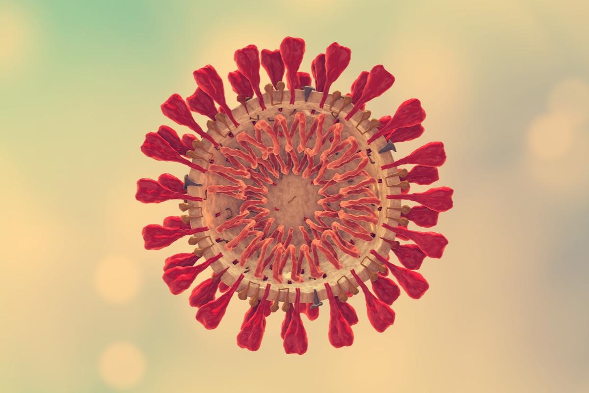
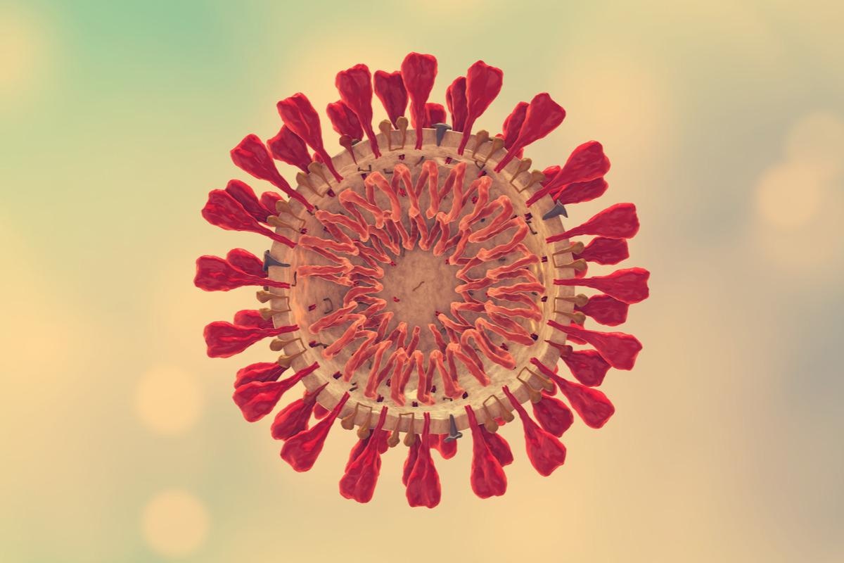
Study: SARS-CoV-2 spike protein–induced cell fusion activates the cGAS-STING pathway and the interferon response. Image Credit: Cinefootage Visuals / Shutterstock.com
Background
A previous study on individuals who died of COVID-19 reported three hallmarks of critical SARS-CoV-2 infection which included the presence of massive lung thrombosis, extensive alveolar damage, and syncytial dysmorphic pneumocytes. Syncytia, which arises due to cell fusion, has also been reported in SARS-CoV-2–infected macaques and in vitro infections. Thus, cell fusion plays an important role in SARS-CoV-2 pathogenicity.
The SARS-CoV-2 spike (S) protein is responsible for triggering cell fusion. Following cleavage of the S protein, it engages with the cell surface angiotensin-converting enzyme 2 (ACE2) receptor for the fusion of the viral membrane with the plasma membrane of the host cell. Thereafter, viral ribonucleic acid (RNA) is released into the host cell cytoplasm for replication.
SARS-CoV-2 uses multiple mechanisms to suppress the production of interferons (IFNs) during the early stages of infection. Several studies have shown that SARS-CoV-2 infection results in the production of low amounts of type I IFNs and high amounts of inflammatory cytokines, while others have shown that this infection can also lead to the production of high amounts of IFNs. This suggests that cells can sense SARS-CoV-2 infection and mount an IFN response, despite the virus evading certain innate immune pathways.
Individuals infected with SARS-CoV-2 can benefit from protection by IFNs, especially type I and type III IFNs. However, prolonged production of IFN can lead to impairment of lung epithelial regeneration.
The production of type I and type III IFNs is induced by the recognition of pathogen-associated molecular patterns by pattern recognition receptors (PRRs). As an RNA virus, SARS-CoV-2 is detected by viral RNA sensors melanoma differentiation-associated protein 5 (MDA5) and retinoic acid-inducible gene I (RIG-I) in infected cells.
A new Science Signaling study analyzes how the activation of DNA sensor protein cytosolic cyclic GMP–AMP synthase (cGAS) and its downstream effector stimulator of interferon genes (STING) can induce a type I IFN response due to SARS-CoV-2 infection.
About the study
The current study involved S protein-mediated cell fusion assays, where target cells were loaded onto the donor cells in a 1:1 ratio. Thereafter, S-protein mediated cell fusion and IFN production were inhibited using inhibitory agents such as the furin inhibitor decanoyl-RVKR-chloromethyl ketone (RVKR) or the lysosomotropic inhibitor bafilomycin A1 (BaflA1).
Sequencing was performed using purified viral RNA followed by RNA-seq data analysis and reverse transcription-quantitative polymerase chain reaction (RT-qPCR) analysis. This was followed by reporter assays, immunofluorescence analysis, western blotting analysis and imaging, as well as 2′3′-cGAMP enzyme-linked immunosorbent assay (ELISA). Replication-competent vesicular stomatitis virus (VSV)–SARS-CoV-2 was used to confirm the effect of S protein expression in fused cells.
Study findings
The S protein was found to alter cellular transcriptomes and induce the transcription of IFNβ and tumor necrosis factor α (TNF-α), as well as IFN-stimulated genes (ISGs) including ACE2 and IFN-induced protein with tetratricopeptide repeats 2 (IFIT2) in fused cells. The S protein was also found to increase phosphorylation of the p65 subunit of nuclear factor κB (NF-κB).
Furthermore, an increase in the nuclear translocation of the transcription factor IFN regulatory factor 3 (IRF3) was reported, along with a smaller ACE2 after cell fusion. The S protein was found to induce the type I IFN response in a time-dependent manner.
S protein mutants with an altered S1/S2 site or an altered S2′ site were resistant to cleavage by proteases. However, the SARS-CoV S protein and S mutants with an altered S1/S2 site could generate modest syncytia, while mutants with an altered S2′ site were unable to induce fusion. Notably, the activation of innate immune responses could not occur with the two S mutants.
Stimulating the expression of IFNβ and the phosphorylation of IRF3 and STING required the expression of both cGAS and STING. The S protein was found to increase the production of 2′3′-cGAMP in cells expressing cGAS and STING.
Knockdown of endogenous cGAS or STING was found to decrease IFN expression, as well as the phosphorylation of IRF3 and STING. Additionally, the activation of cGAS-STING was dependent on cleavage of the S protein and did not occur in S mutants where cleavage cannot take place.
Cell-cell fusion led to increased expression of 429 genes and decreased expression of 382 genes. Most of the increased genes were associated with cell senescence and DNA damage responses (DDRs). Additionally, the presence of micronuclei was observed in S-induced fusion cells but not in fusion cells expressing mutant S proteins.
Notably, cGAS was localized in the micronuclei in fused cells and was associated with the activation of IRF3. S protein expression and syncytia formation of infected cells was confirmed using VSV–SARS-CoV-2, which carried the SARS-CoV-2 S protein. Additionally, cGAS activation and DNA damage were observed during infection with VSV-SARS-CoV-2.
Conclusions
Taken together, the current study suggests that infection with SARS-CoV-2 leads to the activation of cGAS and STING due to micronuclei formation in the fused infected cell. Both cGAS and STING can subsequently activate IFNs and cytokines, which worsens disease severity by eventually causing damage to the lung epithelial cells.
- Liu, X., Wei, L., Xu, F., et al. (2022). SARS-CoV-2 spike protein–induced cell fusion activates the cGAS-STING pathway and the interferon response. Science Signaling. doi:10.1126/scisignal.abg8744. https://www.science.org/doi/10.1126/scisignal.abg8744
Posted in: Molecular & Structural Biology | Cell Biology | Medical Science News | Medical Research News | Disease/Infection News
Tags: ACE2, Angiotensin, Angiotensin-Converting Enzyme 2, Assay, Cell, Coronavirus, Coronavirus Disease COVID-19, covid-19, Cytokines, Cytoplasm, DNA, DNA Damage, Enzyme, Gene, Genes, Imaging, in vitro, Interferon, Interferons, Melanoma, Membrane, Necrosis, Pandemic, Pathogen, Phosphorylation, Polymerase, Polymerase Chain Reaction, Protein, Protein Expression, Receptor, Respiratory, Retinoic Acid, Ribonucleic Acid, RNA, SARS, SARS-CoV-2, Severe Acute Respiratory, Severe Acute Respiratory Syndrome, Spike Protein, Stomatitis, Syndrome, Thrombosis, Transcription, Tumor, Tumor Necrosis Factor, Virus

Written by
Suchandrima Bhowmik
Suchandrima has a Bachelor of Science (B.Sc.) degree in Microbiology and a Master of Science (M.Sc.) degree in Microbiology from the University of Calcutta, India. The study of health and diseases was always very important to her. In addition to Microbiology, she also gained extensive knowledge in Biochemistry, Immunology, Medical Microbiology, Metabolism, and Biotechnology as part of her master's degree.
Source: Read Full Article
