Reduced-dose computed tomography (CT) can safely be used to detect lung metastasis in pediatric patients with various cancers without sacrificing diagnostic accuracy, a study published in the American Journal of Roentgenology concludes.
The study, in which 92% of lung nodules in pediatric patients were detected with reduced-dose CT (0.3 mSv mean effective dose), involved 78 children (44 males) with a mean age of 15 who had diagnoses such as Ewing sarcoma and osteosarcoma. Patients first underwent standard chest CT (1.8 mSv) followed by a chest CT with an 83% dose decrease. A total of 45 patients (58%) had 162 total lung nodules with a mean size of 3.4 mm.
Three radiologists blind-reviewed the CT examinations, and one radiologist conducted a subsequent review to match lung nodules between standard- and reduced-dose CT examinations. The sensitivity of reduced-dose CT for nodules ranged from 63%-77%, and the specificity ranged from 80%-90% across the three radiologists.

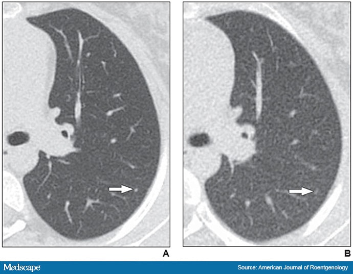
Axial reformatted clinical (1.083 mSv) and reduced-dose (0.318 mSv) CT images from a 17-year-old girl with osteosarcoma. A 2 mm left lower lobe nodule is clearly visible in the left lower lobe on the clinical CT image (arrow in A). The same nodule is vaguely apparent on the reduced-dose CT image (arrow in B), classified as present but poorly visible.
The results point to the potential for increasing the use of reduced-dose CT, according to Andrew Trout, MD, the study’s senior author and an associate professor of radiology and pediatrics at Cincinnati Children’s Hospital, Cincinnati, Ohio.
“In pediatric radiology, we try to reduce dose as it relates to radiation, but we are also trying to balance optimizing the dose with optimizing image quality,” explained Trout. “We don’t want to reduce the dose so low that the exam is not interpretable.”
Trout noted that in clinical practice patients and even clinicians will sometimes choose a chest x-ray over a chest CT because of concerns of exposure to radiation. “There is some hesitancy in some areas to use CT,” he said. “This [reduced dose CT] gives us another tool where we can say that this is about the [same] dose as just a couple of chest x-rays. Patients, parents, and even providers have an idea in their mind that CT is bad because it is a high-dose examination. They sometimes choose an x-ray over a CT scan because of concerns of the radiation dose associated with standard CT.”
Trout explained he and co-investigators chose lung nodules because they are very challenging to identify. “They are very small in size (1 to 2 mm),” said Trout. “If we can prove it in this case, that opens the door to wider use of it in cases where it is appropriate.”
One population that would benefit from reduced-dose CT is children with genetic disorders that make them particularly susceptible to radiation exposure. Another population of patients suitable for reduced-dose CT examinations would be follow-up cases.
“There are some situations where the follow-up examinations have switched over to chest x-ray,” said Trout. “I know that as a radiologist, I can’t detect lung nodules from a chest x-ray as well as I can from a CT scan.”
Trout noted the study is limited by its sample size, the fact that it is a single-center investigation, and that it did not completely reflect “real-world” practice where sedation may have to be used in pediatric patients. He underlined that the study findings do not support the notion that reduced-dose CT can be employed to detect soft tissues in the center of the chest.
“While it may work for lung nodules, we don’t know if it will work for soft tissue lesions in the center of the chest,” said Trout. “The dose reduction will create more difficulties for reading soft tissues in the center of the chest such as the heart. The loss of image quality will be greater.”
Daria Manos, MD, a professor of diagnostic radiology at Dalhousie University in Halifax, Canada, and president of the Canadian Society of Thoracic Radiology, described the study as very carefully designed and one that will be practice-changing.
“They have shown that when the purpose of a CT is to look for lung nodules, we can safely reduce radiation dose,” said Manos, who was not involved in the study. “This is a beautiful example of how we should be further reducing our radiation dose, particularly in children. All of us should be looking at our CT protocols and trying to modify our techniques to reduce the radiation dose as much as we can.”
The use of ultra low-dose CT will decrease cumulative exposure to radiation over the long-term, said Manos. “It’s important to remember that these pediatric oncology patients will often have many CT scans over their lifetime,” she said. “Every reduction in radiation is impactful.”
Manos and Trout have disclosed no relevant financial relationships.
AJR Am J Roentgenol. Published online July 7, 2021. Abstract
For more news, follow Medscape on Facebook, Twitter, Instagram, YouTube, and LinkedIn
Source: Read Full Article
