
Marburg virus is a deadly pathogen that causes Marburg disease a severe viral hemorrhagic fever, named after the city in Germany, where the first outbreak occurred in 1967.
The virus has had several outbreaks across the world since then, with much research focus on its structure and transmission method due to its unusual nature.
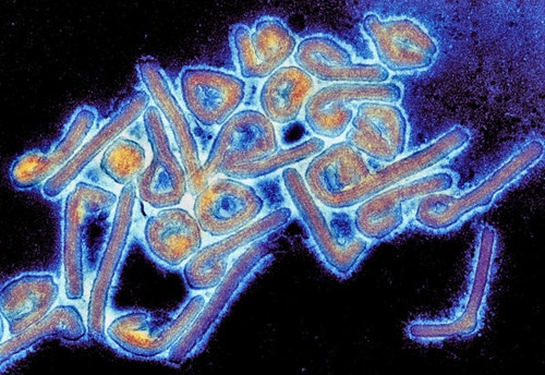
"Colorized Marburg virus particles viewed with a transmission electron microscope" by Microbe World is licensed under CC BY-NC-SA 2.0
Image Credit: Mauro Rodrigues/Shutterstock.com
Research shows that the virus has been found in tears and semen, in addition to the liver even weeks or months after symptoms appear. However, research suggests that transmission of Marburg virus from an infected human is generally unlikely, except when in close contact without protective equipment such as during treatment without barrier nursing or certain burial practices.
Replication cycle
Studying the replication cycle of the Marburg virus is done stepwise, where versions of the virus that lack certain elements to stop them from becoming dangerous are used. While this offers insight into the entry, replication, and budding, they lack the morphological features and protein composition of real Marburg viral particles.
In general, Marburg virus entry into infected cells consists of attachment, endocytosis, and fusion. Both attachment and fusion are mediated by Marburg virus glycoprotein, where it binds to different types of C-type lectins or TAM receptor protein kinases. The method by which endocytosis occurs is not well understood but might occur similarly to the ebola virus, to which it is related.
Transcription and replication of the viral RNA genome occur after the nucleocapsid is released into the infected cell. Seven monocistronic mRNAs are transcribed and encapsidated by the nucleocapsid proteins.
The new nucleocapsids are subsequently recruited to sites where virus budding occurs. Glycoproteins are recruited to the budding sites, where VP40 induces the formation and release of filamentous virus-like particles.
Sources
- Brauburger, K. et al. (2012). Forty-five years of Marburg virus research. Viruses. https://doi.org/10.3390/v4101878
- Towner, J.S. et al. (2006). Marburgvirus genomics and association with a large hemorrhagic fever outbreak in Angola. Journal of Virology. https://doi.org/10.1128/JVI.00069-06
- Brainard, J. et al. (2015). Risk factors for transmission of ebola or Marburg virus disease: a systematic review and meta-analysis. International Journal of Epidemiology. https://doi.org/10.1093/ije/dyv307
Last Updated: Apr 1, 2020

Written by
Sara Ryding
Sara is a passionate life sciences writer who specializes in zoology and ornithology. She is currently completing a Ph.D. at Deakin University in Australia which focuses on how the beaks of birds change with global warming.
Source: Read Full Article