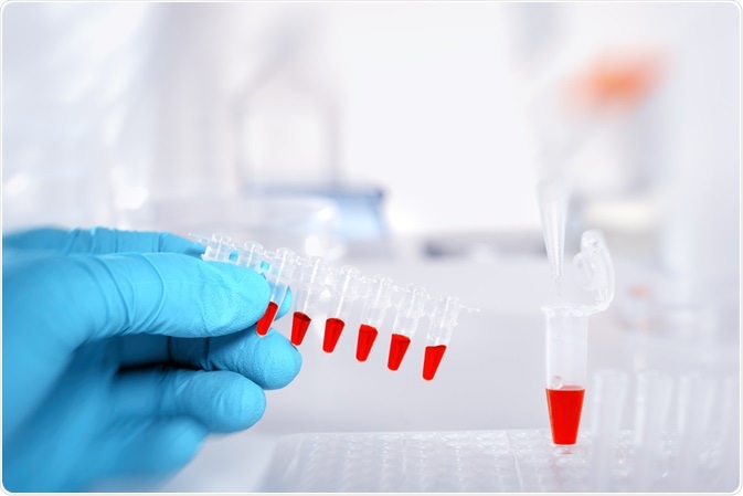
Recent advances in dual-emission ratiometric fluorescence probes have been addressed in a review by researchers at Qingdao University, China. They discuss the applications of biomarker detection and the current state, challenges, and future directions of probes.
 Image Credit: anyaivanova / Shutterstock.com
Biomarker-based detection is essential in the diagnosis and treatment of disease. The ability to detect these biomarkers is dependent on chemo/biosensing and bioimaging. The most attractive are Ratiometric fluorescence (FL) probes that are essential in providing precise, quantitative, visual, and real-time analysis.
FL probes can be categorized into four distinct groups:
- General categories – those used in food sample sensing and live-cell imaging.
- Fabrication methods – those used for tissue and small animal imaging.
- Toxicity distribution – those involved in thin-film visual detection and biological sample sensing.
- Biomarker detection – those used in test paper detection and water sample sensing.
Two general categories for fabricating dual-emission ratiometric FL probes exist – those with one reference signal (target-insensitive) and those with two (dual-emission ratiometric FL). The first category concerns a target-sensitive signal combined with a reference, normalizing the targeted signal via a ratiometric scale.
These show a high signal-to-noise ratio and in the context of chemo/biosensing and bioimaging applications, demonstrate improved accuracy, sensitivity, and reproducibility. Their characteristics, mechanisms, and analytical performances vary depending on the reference and response signal used, targets, and transducer (means of receiving and responding to signals) mechanisms.
In the case of ratiometric fluorescence with two reversible signal changes, two signals have the capacity to induce the increase of one signal (on), together with the decrease of the other (off). Typically, probes have two or more signal outputs to generate an emission and upon binding to targets, produce the reversible variation of 2+ signals – enabling versatility.
Alternatively, a single stimuli-responsive probe is used, which, upon target binding, results in the disappearance of one signal along with the emergence of a new one. Relative to the target-driven reversible changes of different signals approach, a higher signal-to-background ratio is possible. As such, the signaling response is higher ad interference from target-dependent factors is avoided.
In the instance of fabrication methods of ratiometric FL probes for biomarker detection, three main categories exist. These include nanoparticle- or organic dye-embedded probes with dual-emission FL, nanoparticles (NPs), and organic dyes dual-embedded probes.
Fabrication refers to the process of embedding probes into carriers (silica, polymers, liposomes, MOFs, nanogels, etc.), which is achieved via polymerization reactions. Alternatively, physical absorption and chemical conjugation methods can be used on the surface of non-luminescent carriers. The process of fabrication has some limitations, however.
Namely, NPs dyes are in some way compromised i.e., quenching of the NPs-dyes as a result of polymerization, leaking, phototoxicity, etc. Each method has its own advantage, by combinations are considered to be an effective means of preparing high-quality ratiometric FL probes.
Most promising of these techniques is one involving a core comprised of embedded NPs-dyes followed by growing a shell layer to form a core/shell probe, particularly as their advantages include reduced self-quenching and simplicity of design. The authors predict that future studies will explore methods to improve fabrication – focussing on lowering cost, simplifying synthesis, and improved specific and sensitive signal responses to targets.
Different probes have different applications, and these depend on detection mechanisms and context. These range from chemo/biosensing in biological, food, and water samples to bioimaging in cells, tissues, and bodies.
Outside of detection, ratiometric probes can be used in an analytical context where sensitive and selective detection of biomarkers combined with rapid testing is prioritized. Analytical performance is dependent on the efficiency of luminescence, which has not yet been investigated, yet is promising due to their content-independent luminescence.
The production of biomarker detection mechanisms involves immobilizing or dripping ratiometric FL probes onto the surface of solid substrates to produce filter- based sensing platforms – predicted to be an area of significant study in the future, as its flexibility enables intelligent detection of biomarkers. Another future direction is the determination of multiple biomarker detection mechanisms, crucial for accurate early cancer diagnosis and treatment.
With regards to imaging, 3D imaging is currently a powerful means of disease diagnostics; it images real-time in vivo biomarker activity, and the low toxicities and high biocompatibilities of ratiometric probes afford promising biosensing and bioimaging applications in the future.
The authors note that future studies will determine design schemes and best fabrication procedures for the ratiometric FL probe. The improvement in such probes is predicted to offer opportunities in new areas – including flexible and wearable optical devices and advance luminescent detectors.
Acknowledgments
This work was financially supported by the National Natural Science Foundation of China, the Taishan Scholar Program of Shandong, Natural Science Foundation of Shandong, and the Source Innovation Plan Application Basic Research Project of Qingdao
Source
Gui, R. et al. Recent advances in dual-emission ratiometric fluorescence probes for chemo/biosensing and bioimaging of biomarkers. doi: https://doi.org/10.1016/j.ccr.2019.01.004 (2019)
Further Reading
- All Fluorescence Content
- What is Fluorescence Spectroscopy?
- How do Epifluorescence Microscopes Work?
- GFP-tagging in Fluorescence Microscopy
- Fluorescence Quenching
Last Updated: Nov 20, 2019

Written by
Hidaya Aliouche
Hidaya is a science communications enthusiast who has recently graduated and is embarking on a career in the science and medical copywriting. She has a B.Sc. in Biochemistry from The University of Manchester. She is passionate about writing and is particularly interested in microbiology, immunology, and biochemistry.
Source: Read Full Article
